What is Microscopic Digitization?
Conventionally digital images of a prepared slide can be taken during observation under a light microscope at different magnification of objective lens with a digital camera mounted on the microscope. Digital data of single image taken from a limited visual field at a fixed magnification are collected and viewed photo by photo on a computer system like using an album.
After introduction of whole slide imaging (WSI) technology, it has been widely used in pathology and research for creating a single, high-resolution digital file of the entire slide for viewing, archiving, and sharing. In brief, an automated system moves a standard microscope slide under a camera, capturing many high-resolution images (fields of view) that are then stitched together by software.
Our Digital Slides in Plug-and-Play Mode
Up to today, there are more than dozen manufacturers using this WSI technology to produce hardware and software commercially available for clinical and research institutions. Meanwhile, there are few web sites, supported by schools and hospitals, providing free accessibility to use their digital slides from Cloud. However, most Cloud services are based on paid subscription.
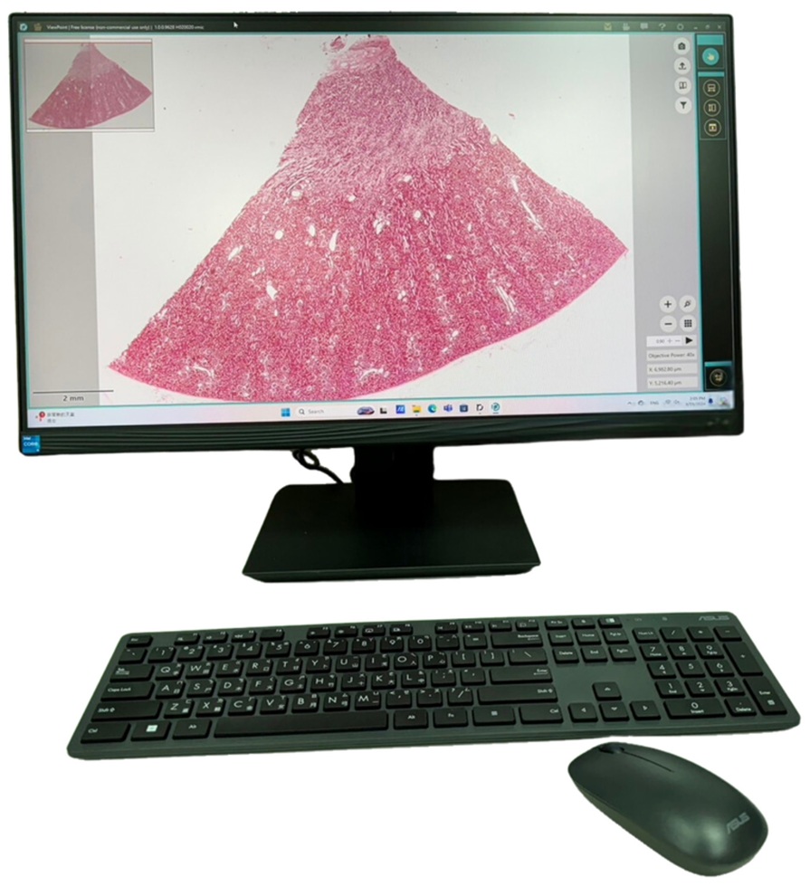
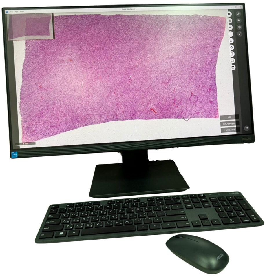
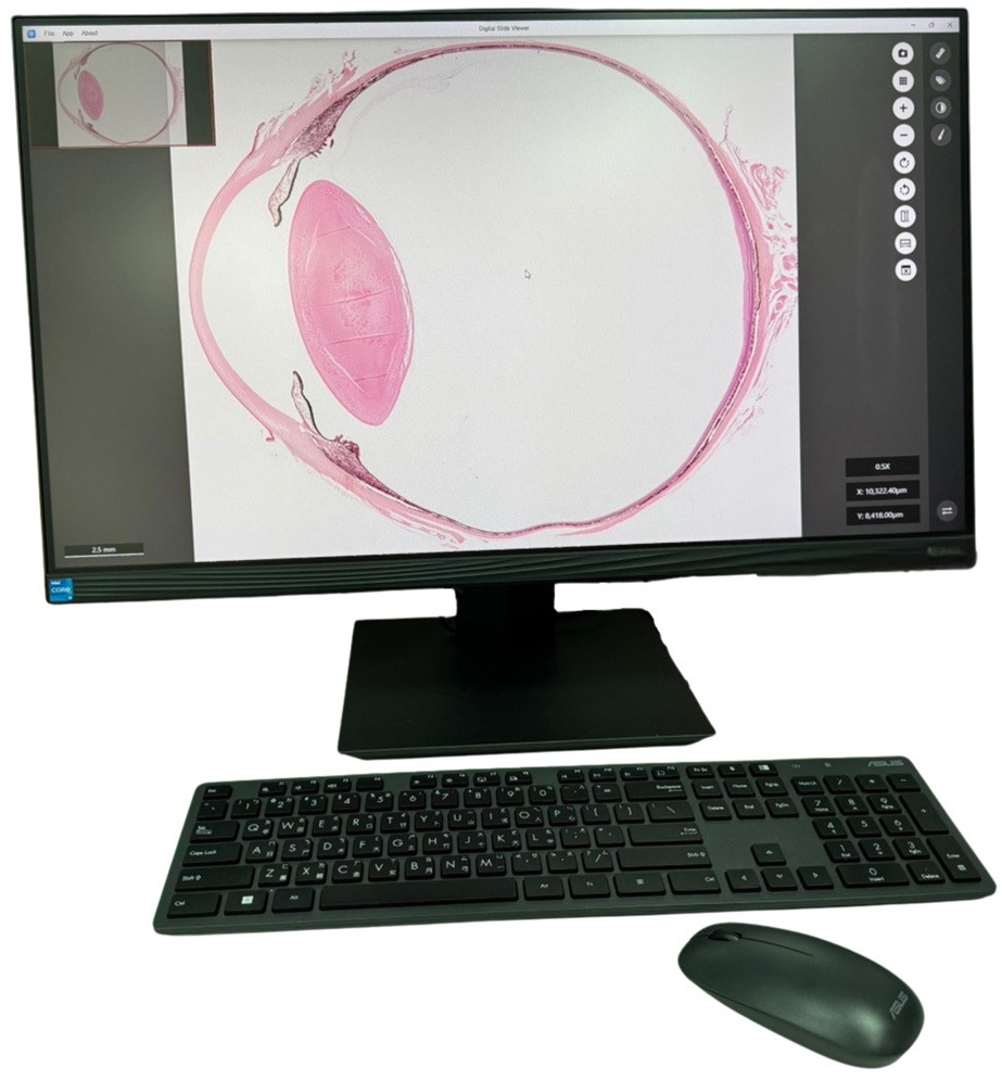
We are the first company providing digital slides loaded in a USB for just plug and play. Installation is not required. There is no need for cloud support. It can be always run without Wi-Fi. There is a three-year warranty on the hardware. An unlimited warranty is provided on the data and software. The Guide document will teach you how to find desired structures on each digital slide at different magnifications. Our own designed DS Viewer enables you to view, rotate, zoom in and out, navigate within thumbnails, delineate regions of interest, measure structures on screen, annotate the structure, and capture to save images for further study.
Currently there are four kinds of digital slides available:
1. Digital Slides for Medical Histology (DSMH)

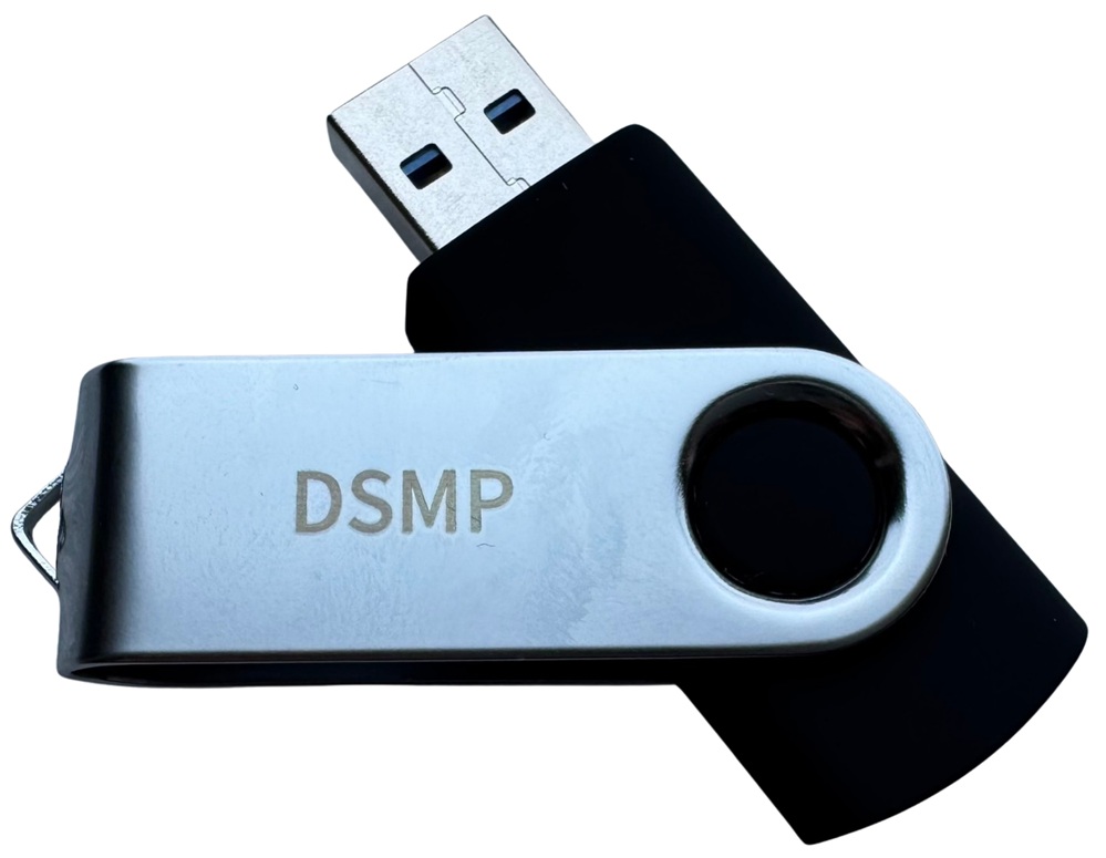
2. Digital Slides for Medical Pathology (DSMP)
3. Digital Slides for Oral Histology and Oral Pathology (OHOP)

4. Digital Slides for Animal Histology (DSAH)

For Users of DSMH, DSMP, OHOP, and DSAH
1. Purchasing any each set of Ginkgomed digital slides will be offered microscopic digitization service of your own 10 prepared slides for free.
2. You need to select your own prepared slides through microscopic observation to ensure the slides have the good quality suitable for creating digital images with high resolution.
3. Once the prepared slides have been sent for digitization, they cannot be replaced due to imperfect quality of the slides.
4. You need to deliver the prepared slides to the location of our distributor where you purchased Ginkgomed digital slides. The distributor will forward the slides to Ginkgomed at your own expense.
5. Your prepared slides will be returned with a USB containing the digitized image data and the Digital Slide Viewer Version 1.5 to you through the distributor.
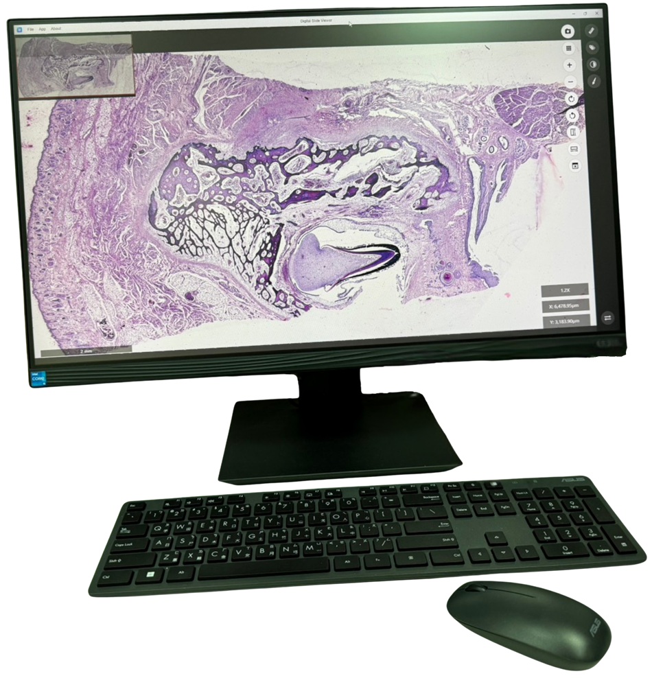
New Features of the Digital Slide Viewer Version 1.5
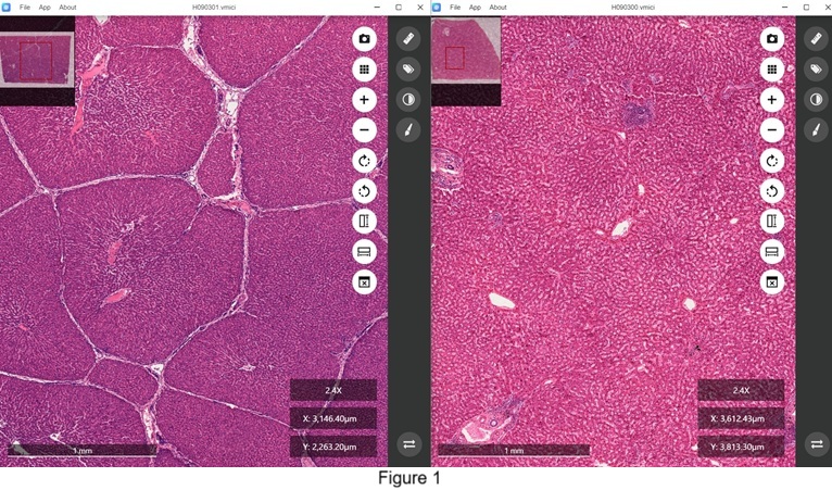
It now allows users to open various image data files in separate windows, enabling independent operation. This enhancement facilitates the concurrent observation and comparison of tissue structures across multiple microscopic sections. One significant application is the comparison of structural differences between human liver and swine liver. As illustrated in Figure 1, the human liver lacks distinct boundaries between hepatic lobules (right), whereas the swine liver is characterized by clearly defined hepatic lobules (left).
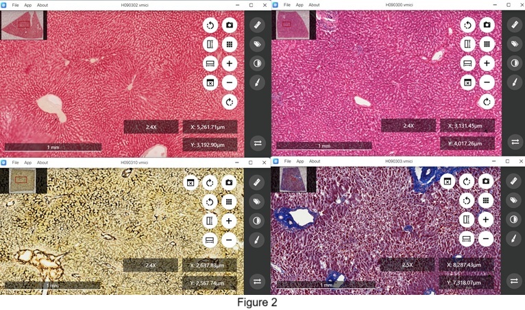
Additionally, this upgraded functionality can be utilized to examine tissue sections from the same source subjected to different treatments, as demonstrated in Figure 2. Human liver sections are analyzed after various staining techniques: hematoxylin-eosin staining reveals cellular organization (upper right), Mallory staining highlights collagenous components (bottom right), PAS staining indicates the presence of glycogen within hepatic cells (upper left), and silver impregnation shows the distribution of reticular fibers in hepatic tissue (bottom left).
Moreover, this tool is invaluable for distinguishing between normal and diseased tissues, as shown in Figure 3. The right window displays a tissue section from a normal human liver, while the left window presents a section from a human liver affected by cirrhosis, with both sections stained using hematoxylin-eosin.

Detailed demonstration of using Ginkgomed Digital Slide Viewer Version 1.5 can be viewed from YouTube at:
For Institutions Not Using DSMH, DSMP, OHOP, and DSAH
Paid service of microscopic digitization is also available for those who are not using Ginkgomed Digital Slides.
Please contact for further information: info@ginkgomed.com.tw