Our Digital Slide Viewer has been upgraded to be capable of opening different image data files in individual windows and operating independently.
User will be able to observe and compare tissue structures of two and even more microscopic sections concurrently.
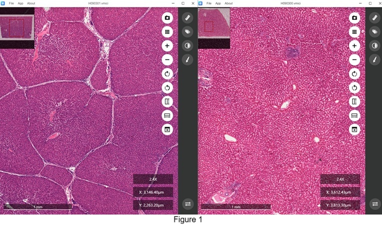
One of the advantages is to compare structural difference between human liver and swine liver as shown in Figure 1.
Human liver does not have apparent boundaries between hepatic lobules (right) while swine liver is composed of hepatic lobules with clear cut boundaries (left).
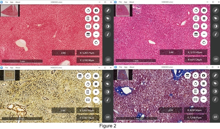
The upgraded function of observing and comparing different microscopic sections concurrently can also apply on viewing tissue sections of the same source with different treatment as shown in Figure 2. Human liver sections are observed after
hematoxylin-eosin staining to show cellular organization (upper right), Mallory staining to reveal collagenous components (bottom right), PAS staining to indicate existence of glycogen inside the hepatic cells (upper left), and sliver impregnation to shown distribution of reticular fibers in the hepatic tissue (bottom left).
It definitely can be applied to distinguish differences between normal and diseased tissues as shown in Figure 3. In this figure, the right window shows tissue section taken from normal human liver while the left window shows tissue section taken from human liver with cirrhosis. Both sections are stained with hematoxylin-eosin.
