

The software is contained in a dongle protected SSD external drive.
It is the first 3D anatomy software using real human dissected specimens in the world. The 3D presentation is designed from more than 300 real human specimens, enough for learning key structures of the entire human body. These specimens are well-dissected and preserved in a good condition for creating 3D visualization.
There are six body regions to be studied:
- HEAD AND NECK.
- TORSO AND BACK.
- THORAX.
- PECTORAL GIRDLE AND UPPER LIMB.
- ABDOMEN AND PELVIS.
- PELVIC GIRDLE AND LOWER LIMB.

The following demonstration will give you a quick apprentice of how it can help in learning anatomy. For an example, to study the anatomy of the HEAD AND NECK, click the “Intracranial region”, 12 brain specimens will be shown on the screen.

Click to select “Arteries of the brain”. The image of the specimen appears on the center of the screen with structure terms shown on the left.

Click any artery shown on the specimen image, the term of the artery appears at the bottom with the pronunciation.

Click the term at the bottom, a description of the anatomical structure will be shown.

The desired structure can also be localized by clicking the structure term on the left.

The image of the specimen can be zoomed in or zoomed out by rolling mouse wheel up or down.

By holding the left key, the image of the specimen can be turned around at any angle on the screen.

By holding the mouse wheel, the image of the specimen can be moved around on the screen under the control of mouse movement.

Clicking the “Background” on the right menu bar, you may change color of the background.

Click the “Brush”, you may do colored marks on the screen.

You also can do “Screen shot” to save the image.


The saved image can be used in Word or Power Point files to create your own content for research paper, lecture, or making an exam.
Other control commands:
Lock: All control commands will be locked.
Previous: Click to be back to the previous one.
Next: Click to move forward to the next specimen.
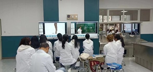
This 3D Anatomy Software can be used with a window-based PC or notebook for professor’s teaching preparation or personal study. It also can be used with a touch screen for anatomical lectures in the class, and even used as a dissection guide for a big group of students in the anatomy laboratory.

An example of 3D Anatomy Installation in a digital anatomy laboratory is shown below :


A “Classroom Practice” has been added into the content of the he newly upgraded version 1.1 as shown below :


There are thousands of quiz questions designed to test users’ learning of human anatomy. If the question is answered correctly, the answer will be shown with a green background. If the answer is wrong, the answer will be marked with a red background.
A correct answer with a green background also appears. In this mode, it will help students learn anatomy efficiently.